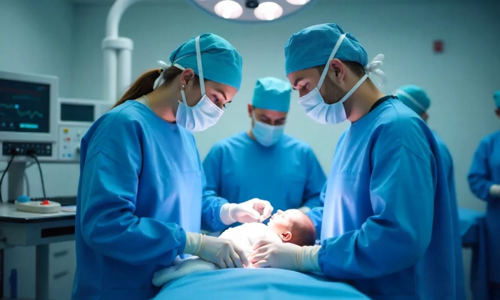
Gastroshiza: Causes, Symptoms, and Treatment Options Explained
When parents first hear about gastroshiza (also known as gastroschisis), it can feel overwhelming and frightening. This congenital anomaly affects about 1 in every 2,400 newborns, where intestines and sometimes other bowel structures develop outside the abdominal wall. While this newborn intestinal condition sounds scary, modern pediatric surgery and neonatal intensive care have dramatically improved outcomes for these little ones.
What is Gastroshiza?
Gastroshiza (gastroschisis) is an abdominal wall defect where a baby’s abdominal wall doesn’t form completely during pregnancy. This creates an opening, usually to the right of the umbilical cord, allowing intestines and sometimes other organs to develop outside the body with organ exposure in babies occurring throughout development.
This congenital anomaly develops early in pregnancy, typically between the 6th and 10th weeks. During this crucial time, the abdominal muscles should undergo proper abdominal wall closure around the developing organs. When this process doesn’t happen correctly, this abdominal wall defect occurs.
Unlike other abdominal wall defects, the intestines in this condition are not covered by a protective sac. They float freely in the amniotic fluid throughout pregnancy, which can cause irritation and damage. This exposure is what makes gastroschisis different from similar conditions like omphalocele, where organs are covered by a membrane.
Understanding this difference is important because birth defect treatment approaches vary significantly between these conditions. While both require surgical repair for gastroschisis and omphalocele respectively, the timing and methods differ based on whether organs are protected or exposed.
Comparison Table: Gastroshiza vs. Omphalocele
| Feature | Gastroshiza | Omphalocele |
|---|---|---|
| Organ covering | No protective sac | Covered by membrane |
| Location | Right of belly button | At belly button |
| Associated conditions | Usually isolated | Often with other defects |
| Surgery timing | Soon after birth | May be delayed |
Causes of Gastroschisis
The causes of gastroschisis remain largely unknown to medical experts. However, research has identified several genetic and environmental risk factors that may increase the likelihood of this congenital abdominal wall defect developing during pregnancy.
Young maternal age stands out as the strongest risk factor. According to Dr. Feldkamp, an expert in gastroschisis epidemiology, “Young maternal age is the strongest and most consistent risk factor.” Mothers under 20 years old have significantly higher rates of infants born with this condition.
Smoking during pregnancy also increases the risk. Studies show that mothers who smoke have a higher chance of having a baby with gastroschisis compared to non-smokers. Other genetic and environmental risk factors like poor nutrition, certain medications, and exposure to environmental toxins may also play a role.
Genetic factors appear less significant in this condition compared to other birth defects. Most cases occur randomly without a family history of the condition. This means parents shouldn’t blame themselves, as this congenital anomaly typically isn’t something that runs in families or results from specific actions during pregnancy.
Does diet cause this condition?
One common myth suggests that poor diet directly causes this birth defect. While good nutrition supports healthy pregnancy, there’s no evidence that specific foods cause this condition. However, maintaining a healthy diet with proper vitamins and minerals, especially folic acid, supports overall fetal development and may reduce various birth defect risks.
Recent data from the CDC shows that these rates in the US have remained relatively stable, affecting about 1 in every 2,400 to 2,500 births. Interestingly, some research indicates a slight decline in cases from 2016 to 2022, though experts continue studying these trends.
Recognizing Symptoms of Gastroschisis in Infants
This newborn intestinal condition is immediately visible at birth, making diagnosis of gastroschisis before birth and after delivery straightforward for medical teams. The most obvious sign is intestines protruding through an opening in the baby’s abdominal wall, typically on the right side of the umbilical cord.
These exposed intestines may appear swollen, thick, or discolored due to prolonged exposure to amniotic fluid during pregnancy. The intestinal walls often look irritated or inflamed, which is normal given their exposure throughout development.
Newborns with this condition may face several risks and complications in gastroschisis beyond the visible defect. Feeding difficulties are common since the bowel may not function normally at first. Some infants experience trouble digesting milk or formula, requiring special feeding approaches or temporary feeding tubes.
Growth delays can occur, especially in the first months of life. The exposed intestines lose heat and fluids more easily, making it harder for babies to maintain proper nutrition and growth patterns. Infection risk is also higher due to the open abdominal cavity.
Can this condition be detected before birth?
Yes, diagnosis of gastroschisis before birth can usually be achieved during routine pregnancy ultrasounds. Most cases are identified during the second trimester, typically between 18 and 22 weeks of pregnancy. The ultrasound clearly shows intestines floating outside the abdominal cavity.
Early detection allows medical teams to plan for delivery and immediate care. Pregnant mothers may need more frequent monitoring and specialized delivery planning. Many hospitals recommend delivery at facilities with neonatal intensive care units and pediatric surgeons readily available.
Checklist for Parents: Signs doctors check during pregnancy and at delivery
- Intestines visible outside abdomen on ultrasound
- Proper fetal growth monitoring
- Amniotic fluid levels
- Signs of intestinal damage or complications
- Heart rate and movement patterns
- Planning for specialized delivery care at facilities with Neonatal Intensive Care Unit (NICU) capabilities
Treatment Options for Gastroschisis
Immediate treatment options for gastroschisis begin the moment a baby with this condition is born. Medical teams quickly cover the exposed intestines with sterile, moist dressings to prevent infection and heat loss. The newborn receives intravenous fluids and antibiotics while preparing for neonatal surgery.
Pediatric surgery is typically required within the first 24 to 48 hours of life. The timing depends on the baby’s overall condition and the extent of the defect. Some infants may need emergency surgery immediately, while others can wait slightly longer for optimal surgical conditions.
Two main approaches exist for surgical repair for gastroschisis. Primary closure involves putting all the intestines back into the abdomen and closing the opening in one operation. This is possible when the abdominal cavity is large enough to accommodate the intestines without creating dangerous pressure.
Staged repair becomes necessary when the intestines are too large or swollen to fit safely back into the abdomen immediately. Pediatric surgeons use a special plastic pouch called a silo to gradually reduce the intestines back into the body over several days or weeks.
Types of surgical repair
The choice between primary closure and staged repair depends on several factors. Neonatologists and pediatric surgeons consider the size of the abdominal opening, how much the intestines have grown outside the body, and the baby’s overall health status.
Recovery from congenital abdominal wall defect typically involves a lengthy stay in the Neonatal Intensive Care Unit (NICU). Babies often need breathing support, specialized feeding, and careful monitoring for complications. The intestines may not work normally at first, requiring patience as digestive function gradually improves.
Given the extensive medical care required, families should consider comprehensive health coverage to manage the significant costs associated with NICU stays, surgical procedures, and long-term follow-up care.
Most babies with this condition cannot eat normally right away. Feeding after birth gastroschisis often requires nutrition through intravenous lines for weeks or months while the intestines heal and begin functioning properly. Gradually, they can start receiving small amounts of breast milk or formula through feeding tubes if necessary.
Living with This Condition: Parental Support for Gastroschisis Care
Bringing home a baby who has undergone surgical repair requires special attention and care. Parents need to monitor the surgical site for signs of infection, ensure proper wound care, and attend regular follow-up appointments with pediatric surgeons and neonatologists.
Feeding challenges often continue after leaving the hospital. Many infants need specialized formulas or continued tube feeding for weeks or months. Working closely with pediatric dietitians and gastroenterologists helps ensure proper nutrition and growth during recovery from congenital abdominal wall defect.
Emotional support is crucial for families navigating this journey. Many parents experience anxiety, guilt, or overwhelming stress when caring for a baby with this abdominal wall condition. Connecting with other families who have faced similar challenges can provide valuable perspective and encouragement.
Support groups, both online and in-person, offer practical advice and emotional comfort. Many children’s hospitals have specialized programs connecting families affected by this condition. Social media groups and foundations dedicated to parental support for gastroschisis care also provide resources and community support.
Can a child live a normal life after surgical repair?
The majority of children with this condition go on to live completely normal, healthy lives. While recovery takes time and patience, most develop normal digestive function and can eat regular foods without restrictions as they grow.
Some children may experience ongoing digestive sensitivities or require occasional medical follow-up. However, these issues rarely prevent them from participating in normal childhood activities, sports, or academics. Many grow up without any lasting effects from their early medical challenges.
Early intervention and consistent medical care significantly improve long-term outcomes. Children who receive proper surgical repair and follow-up care typically reach normal developmental milestones and enjoy the same quality of life as their peers.
Helpful resources for parents:
- Free online parenting support groups
- Hospital-provided care guides and education materials
- Mobile apps for tracking feeding, growth, and medical appointments
- Specialized foundations offering informational resources
- Connection to families with similar experiences
Insert real-life success story: Many parents share inspiring stories of children who overcame these challenges to become active, healthy kids participating in sports, academics, and all normal childhood activities.
Conclusion
This birth condition occurs when babies are born with intestines outside their abdominal wall due to incomplete development during pregnancy. While the exact causes remain unclear, factors like young maternal age and smoking increase the risk.
The condition is easily recognizable at birth and requires immediate surgical intervention. Modern surgical techniques and neonatal care have dramatically improved survival rates and long-term outcomes for affected babies. Most children go on to live completely normal, healthy lives after proper treatment and recovery.
Parents facing this diagnosis should know that support and resources are available throughout the journey. Working with experienced medical teams, connecting with other families, and maintaining hope are essential elements of successful treatment and recovery.
